
The method, pioneered by researchers at the Paul Scherrer Institute (PSI) and the EPFL in Switzerland, could in future be used to detect the onset of breast cancer by highlighting subtle changes in the tissue of the breast.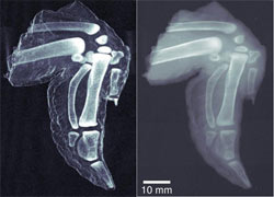
The new method produces dark-field X-ray images, which show the scattering of radiation within materials and provides more detail than ordinary X-rays; as cancer cells scatter radiation differently to healthy tissue, this can be picked up using the new technique.
Professor Franz Pfeiffer says researchers have been working on dark-field X-ray images for many years but until now these images have only been possible using sophisticated crystal optics which are inefficient as they only work for a single X-ray wavelength.
The new process involves taking four separate images, each with the three filters in a slightly different arrangement and software then compares each snapshot to produce a final high-contrast picture.
The resulting images reveal physical details that would normally be invisible.
Pfeiffer say although the process means exposing the subject to a higher total dose of radiation, this can be justified in some circumstances and could be used for some clinical investigations such as mammography.
Pfeiffer's team is currently working on making the process suitable for more powerful X-ray machines and they plan to test it on live animals within the next 12 months.
Researcher Christian David also from PSI, says the new technique opens up the possibility for adapting current imaging equipment to include dark-field imaging and the technique could also be used to diagnose changes in bone density that might signal osteoporosis, as well as plaques in the brains of patients with Alzheimer's disease.
